Description
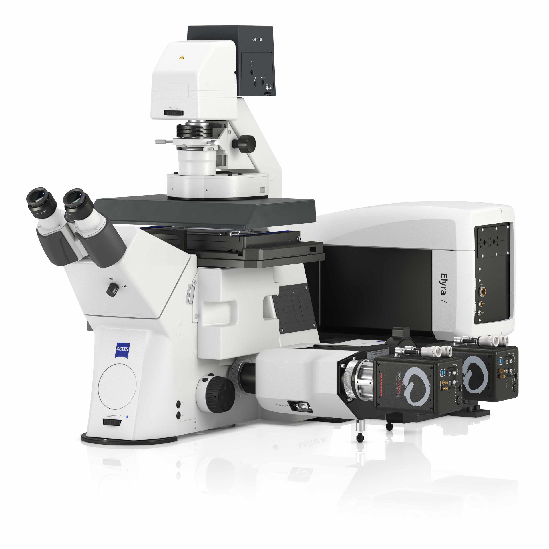
The Confocal Microscope is a state-of-the-art imaging system designed for high-resolution, three-dimensional visualization of cells, tissues, and biomolecular structures. Utilizing laser scanning technology, it provides precise optical sectioning, eliminating background noise and enhancing contrast for detailed analysis in biological and material sciences.
Key Features:
-
- High-Resolution Optical Sectioning:
- Uses laser scanning technology to generate crisp, 3D images with minimal background noise.
- Captures fine details at the subcellular level for precise structural analysis.
- Multi-Channel Fluorescence Detection:
- Simultaneous detection of multiple fluorophores for co-localization studies.
- Advanced spectral unmixing ensures clear separation of overlapping signals.
- Live Cell Imaging & Time-Lapse Capabilities:
- Supports real-time visualization of dynamic cellular processes.
- Environmental control systems maintain optimal temperature, humidity, and CO₂ levels.
- Super-Resolution & High-Speed Scanning:
- Advanced deconvolution algorithms enhance resolution beyond the diffraction limit.
- Fast frame rates enable rapid imaging of cellular events and high-throughput screening.
- 3D Reconstruction & Image Analysis:
- Generates high-fidelity 3D models of cells and tissues.
- Integrated software provides automated quantification, segmentation, and visualization tools.
- Adaptive Optics & Deep Tissue Imaging:
- Corrects for optical aberrations, ensuring consistent image quality.
- Multiphoton and light-sheet options enable deeper tissue penetration with minimal photodamage.
- Automated Workflow & AI-Powered Analysis:
- Automated slide scanning for batch processing of samples.
- AI-driven image recognition accelerates data analysis and feature identification.
- Seamless Data Integration & Connectivity:
- Compatible with cloud-based storage for easy data sharing and collaboration.
- Direct integration with laboratory management systems for streamlined workflows.
- High-Resolution Optical Sectioning:
Applications:
-
- Cell Biology & Neuroscience: High-resolution imaging of live and fixed cells, neuronal networks, and subcellular structures.
- Cancer & Disease Research: Visualization of tumor microenvironments, cell signaling pathways, and molecular interactions.
- Immunofluorescence & Protein Localization: Detailed mapping of protein expression and co-localization studies.
- Developmental Biology & Stem Cell Research: Time-lapse imaging of embryonic development and differentiation.
- Material Science & Nanotechnology: Surface characterization and analysis of polymers, nanomaterials, and bioengineered structures.